Poster Presentation 51st International Society for the Study of the Lumbar Spine Annual Meeting 2025
Data-driven imaging features and their association with lumbar spinal stenosis in pre-surgical patients (#115)
Introduction
Surgery for lumbar spinal stenosis is effective for leg pain and to increase walking distance for most patients, however suboptimal patient selection and individual risk factors may contribute to unfavorable outcomes, especially regarding back pain. This underscores the need for a deeper understanding of the condition and the identification of prognostic factors for outcome. This study aims to provide an objective, data-driven assessment of tissue changes related to stenosis using measurable imaging features. These features could help classify pain types, identify segments at risk of instability, and enhance understanding of degenerative spine changes, ultimately improving treatment strategies. Lumbar segments planned for decompressive surgery were here compared to segments not planned for surgery.
Methods
Data from the multi-center NORDSTEN study, involving 16 hospitals across Norway, were used for this analysis. The study included 344 patients (66.7 ± 8.3 years; 182 men) with lumbar stenosis. A total of 1032 spinal motion segments, consisting of one intervertebral disc (IVD) and two adjacent vertebrae, were examined using T1- and T2-weighted MR images before surgery. The analysis focused on vertebra and discs between L2 and L5. All IVDs and vertebrae were segmented using an in-house AI segmentation tool, and each IVD was further subdivided into five equal subregions, ranging from 1 (anterior) to 5 (posterior) IVD.
For each motion segment, 37 image features calculated from the T1- and T2-weighted MRI scans were tested for significance based on whether the IVD was planned for surgical decompression. Levene’s-test assessed the equality of variances for features involving standard deviation calculations. For the remaining features, the Mann-Whitney U-test was applied. All tests were Bonferroni-adjusted for multiple comparisons, with statistical significance set at p<0.05. Statistically significant features were further analyzed using point-biserial correlation and visualized with boxplots.
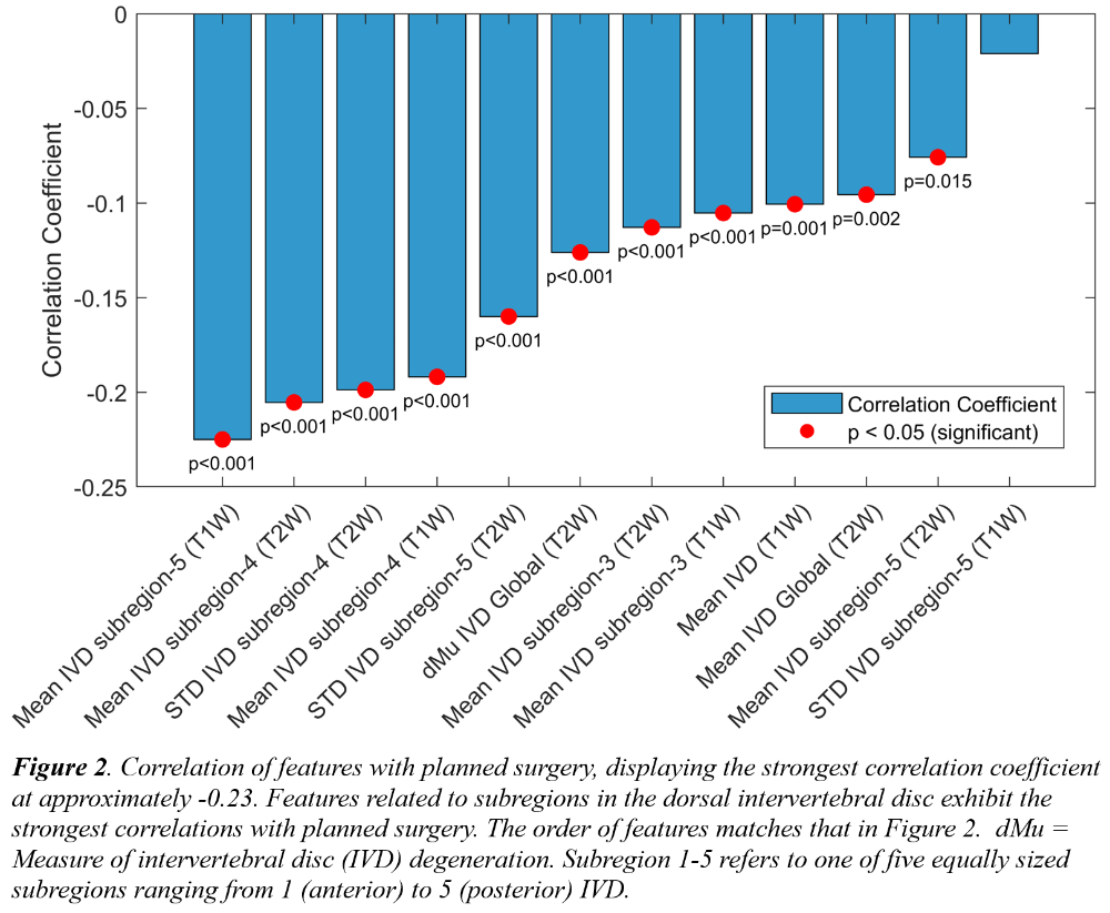
Results
Twelve features were significantly different between segments that were planned for surgical decompression or not (Table 1, Figure 1). Eleven of these features were significantly negatively correlated to planned surgery (Figure 2), i.e., features calculated from segments with stenosis had lower values than those without stenosis. Features related to the dorsal IVD exhibited the strongest correlations.
Discussion
This study highlights the association between imaging features and planned surgery for lumbar stenosis. While vertebral tissue changes show a limited correlation with stenosis, significant correlations were observed for features related to IVDs, especially in their dorsal regions. These findings suggest that the dorsal regions of IVDs tend to be more degenerated in segments with stenosis. Such increased degeneration could result from the dorsal regions being exposed to greater mechanical stress1, which might increase susceptibility to degenerative changes. Additionally, the feature dMu, which quantifies global IVD degeneration2,3, appears to correlate with stenosis, supporting previously known findings that IVDs at levels with stenosis are often more degenerated. These objective features hold promise for future AI-driven multiparameter analyses, enabling synergistic value by combining them to offer new insights into pain-related changes and ultimately improve treatment strategies.
1.Torén L, et al. Spine, 2024
2.Waldenberg C, et al. Eur.SpineJ., 2017
3.Li Z, et al. Eur.SpineJ., 2024