Special Poster Session 51st International Society for the Study of the Lumbar Spine Annual Meeting 2025
Distinctive Features of Upright CT Myelography in Patients with Lumbar Spine Degenerative Diseases (#SP-7d)
Introduction:
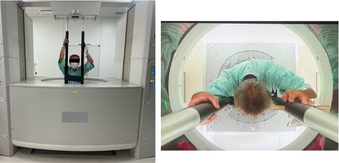
In lumbar spine degenerative diseases (LSDD), gravity plays an essential role in the manifestation of clinical symptoms1. Recently, a full-body upright CT scanner has been developed (Figure 1) 2. The upright CT, when combined with myelography, is anticipated to be an exceptionally modality for understanding the pathology in patients with LSDD. This study aimed to elucidate the distinctive features of upright CTM in LSDD by comparing them with supine magnetic resonance imaging (MRI).
Methods:
We included patients who underwent both supine MRI and upright CTM for LSDD, such as lumbar spinal canal stenosis, lumbar disc herniation, lumbar degenerative spondylolisthesis, lumbar spondylolysis, and lumbar degenerative scoliosis. The dural sac anteroposterior diameter, transverse diameter, and area were measured from L1/2 to L5/S1 on both supine MRI and upright CTM. The following data were collected: age, sex, body mass index (BMI), and spinal sagittal alignment parameters on whole standing spine X-rays, including the sagittal vertical axis (SVA), lumbar lordosis (LL), and pelvic tilt (PT), was evaluated using standing X-ray. In addition, LL was also measured on both supine MRI and upright CTM.
Results:
Data from a total of 110 patients with LSDD were analyzed. The anteroposterior diameter of the dural sac was significantly smaller at L2/3 and L4/5 in the upright CTM, while the transverse diameter was significantly larger at L1/2 and L2/3 in the upright CTM. The dural sac area was significantly larger at the L1/2 level and significantly smaller at the L4/5 level in the upright CTM. A subgroup analysis was performed with 56 patients, whose LL in the upright CTM was greater than in the supine MRI, categorized as Group I, and 54 patients, whose LL in the upright CTM was smaller than in the supine MRI, categorized as Group D. There was no significant difference in age or sex between the two groups, whereas BMI was significantly higher in Group D. LL and SVA in the standing X-ray images were significantly larger in Group I compared to Group D, while PT was significantly smaller in Group I than in Group D. In Group I, the anteroposterior diameter of the dural sac was significantly smaller in upright CTM compared to supine MRI at the L2/3, L3/4, and L4/5 levels. As a result, the dural sac area was significantly smaller at the L3/4 and L4/5 levels. In Group D, no significant differences were observed in the anteroposterior diameter of the dural sac between upright CTM and supine MRI, while the dural sac area was significantly larger in upright CTM than supine MRI at the L1/2 level.
Discussion:
Our results showed that changes in the anteroposterior and transverse diameter and area of the dural sac during the transition from supine to standing vary markedly depending on the intervertebral level. Moreover, it was shown that these parameters are influenced by patterns of change in LL between the standing and supine positions. The use of upright CTM revealed that LSDD patients exhibit diversity in the position-related morphological changes of the dural sac.
- Kreiner DS, Shaffer WO, Baisden JL, et al. An evidence-based clinical guideline for the diagnosis and treatment of degenerative lumbar spinal stenosis (update). Spine J. 2013;13(7):734-43.
- Jinzaki M, Yamada Y, Nagura T, et al. Development of Upright Computed Tomography With Area Detector for Whole-Body Scans: Phantom Study, Efficacy on Workflow, Effect of Gravity on Human Body, and Potential Clinical Impact. Invest Radiol. 2020;55(2):73-83.